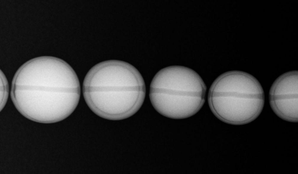
Cultured pearls seen under X-rays
To distinguish natural pearls from cultured pearls, our operators use an X-rays imagery instrument, of which the principle is similar to the one of medical radiography. By observing X-rays images, one can see the inner structure of the pearl.
For the cultured pearls with nuclei shown here, a nucleus is placed inside the mollusc by a human being so that it is covered with a layer of mother-of-pearl, which will more or less thick, depending on how long it has been in the oyster.
The first cultured pearls appeared in Japan and are said to have been created by Kokichi Mikimoto in 1893 using Akoya oysters (Pinctada fucata).
Today, several other species of oysters are used and there are other methods for obtaining cultured pearls, e.g. by "greffons". There are also freshwater cultured pearls, of which China is by far the world's leading producer.
In the X-ray image shown here, a nucleus almost as big as the pearl in size is observed, and is covered with a thin layer of mother-of-pearl, we can assert easily the examined pearl is a cultured pearl.
Do you need this micrograph in full resolution for an article, a DUG dissertation, etc.? Do not hesitate to contact us.